• Skip to main content

College Hill Pilates and Physical Therapy LLC

Pilates Classes in Cincinnati

(513) 445-WELL (9355)
  • Home
  • Physical Therapy
    • Telehealth PT
  • Pilates
    • Pilates Mat Classes
    • Virtual Pilates Classes
  • Strength Training
  • Yoga
  • Tai Chi
  • Blog
  • Schedule

EDS

Feb 01 2022

Experiencing a Catastrophic Shoulder Injury from a Physical Therapist’s Perspective, One Year Later

I haven’t posted an update about my shoulder rehab process since last April, which is when I returned fully back to work. Not really a coincidence!

As of today it has been one year since my surgery to repair my torn labrum and Hill-Sachs lesion. For anyone just now catching this blog, I dislocated my shoulder when I tripped on a sidewalk crack and fell while jogging early in the morning January 14, 2021. I have been working very hard all year to rehab my shoulder. I would say it’s about 95% recovered at this point. I’m hopeful that there is still some improvement to be seen.

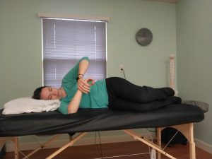
It has been a challenge to regain the mobility and rebuild the strength at the same time. I’m happy to report that my range of motion is nearly 100%. Functional reach behind my back is still a little limited but good enough to fasten a bra behind my back or loop my belt around the back of my waist. My strength is also mostly back to normal. I am able to do all of my work activities, including manual therapy, that I would normally do. I don’t notice many functional limitations at home either and was even able to return to playing tennis with my family this summer.

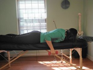
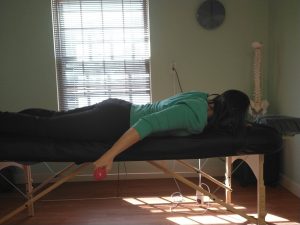
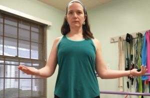
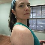
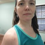
The big issue is still that pesky scapulohumeral rhythm, especially when I reach forward and up overhead.

https://www.collegehillpilatespt.com/wp-content/uploads/2022/02/Shoulder-flexion-and-scaption.mp4

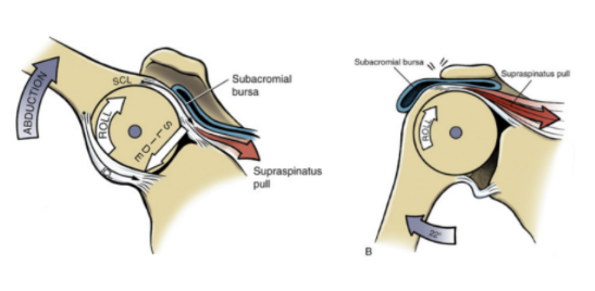
You can see that my scapula still wings a little bit as I raise my arms up and down. This is caused by the continued limited range of motion of the actual ball-and-socket joint of my shoulder. So more of the motion has to come from the shoulder blade moving excessively. This causes pinching of the soft tissue structures in the subacromial space because the humeral head does not glide downward as it rolls upward. As a result, these impinged structures cause pain. This pain is the biggest problem that is yet to resolve. I notice it the most at night because I still wake up several times a night in pain. If I spend too long in any sleeping position, it becomes painful and wakes me up. Happily, I am able to fall back asleep once I shift positions are rearrange myself. But my ultimate goal is to not wake up at night due to pain.

Glenohumeral joint arthrokinematics
On the left we have normal joint arthrokinematics. On the right you can see the pinching above the humeral head (the “ball” part of the joint) that results from altered joint mechanics.

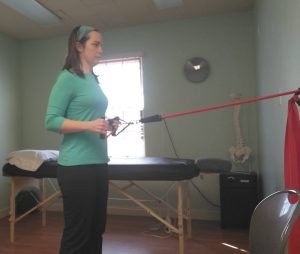
Even though it has been a full year since surgery, I feel like this is still slowly getting better. So there is still more room for improvement. I plan to continue working on loosening up the part of the joint capsule that is still restricting this movement, strengthening the scapular stabilizers, and working on neuromuscular control of the entire pattern. So I’m still hopeful it will get even closer to 100%.

I can’t round out this year of shoulder rehab without mentioning a little bit about my continuing work to recover mentally and emotionally from this injury as well. While tripping on a sidewalk crack might not seem all that traumatic or frightening, it really affected me. I still have anxiety about walking on the sidewalk, especially if it’s dark outside. I haven’t been able to run since this injury, and even watching other people run on the sidewalk gives me a lot of anxiety. (Run on the street, my friends!) I see this a lot with my patients. We develop avoidance behaviors, consciously or subconsciously, because something we did caused us pain. Sometimes it’s something big and traumatic, but sometimes it’s something little like hurting your back when you bent down to pick up a sock. So ever after you have fear that you’ll relive that pain whenever you pick up a sock. Or maybe you felt a tweak in your back and ignored it and a few days later that tweak turned into intense pain down your leg. So now every little tweak you feel sets off alarm bells. I began seeing a mental health counselor a couple of months ago to try to work through some of this because, while running is not my favorite thing to do, I would like to be able to do it if I feel like it without fear or dread. And as a PT I do need to be able to watch my patients run now and then to analyze a faulty gait pattern. I’m hopeful that working through some of these issues on the mental health side of the equation will also help me get to full recovery and prevent this incident from affecting what I can do for any reason, physical or mental.

Nothing is lost of course. This experience has given me more empathy for my patients, though that empathy has probably actually made me a tougher rather than an easier PT! (I’ve been through it, you can do it too!) Feeling the tightness and tension in my own shoulder has helped me understand the way some of the treatments I perform and prescribe for my patients feel, and how they can help. Prior to this injury all of my joints were so loose and hypermobile that I had a hard time understanding what restricted joint mobility feels like. Now I know!

I hope that in writing about my experience with this challenge I’ve helped people have a better understanding of injury, whether their own or someone else’s. I hope that reading this reminds you of the awesome resilience and general coolness of the human body and, of course, of the power of physical therapy!

Written by Mindy Nagel, PT, DPT, OCS · Categorized: EDS, Hypermobility, Physical therapy, Proprioception, Range of motion, Scapulohumeral rhythm, Shoulder, Shoulder injury, Shoulder rehab, Shoulder surgery

Apr 15 2021

Experiencing a Catastrophic Shoulder Injury from a Physical Therapist’s Perspective, Part 14 — Back to Work!

Busy, busy, busy!

Yesterday was the 3 month anniversary of my shoulder dislocation caused by tripping on a sidewalk crack while jogging. The strange thing is that even after 3 months I still feel detached from this injury, like I’m watching someone else go through it and working on it as if I were my patient. When I talk about the injury and the recovery process it feels like I’m talking about someone else. I’ve read that this can happen as a result of trauma. But I think it also has to do with the fact that I am a physical therapist and therefore my natural inclination is to look at an injury objectively with an outside observer’s perspective.

I haven’t written a blog entry about my shoulder injury recovery in a while because I have been so busy with the re-opening of my office! It didn’t take long to get back up to a full schedule of patients and keeping up with that is just a little challenging with my shoulder not being 100%. It’s a common challenge that as we recover we start to add our usual activities back into our lives and the time we have to devote to rehab, rest, and recovery starts to get chipped away. So I’m working really hard to balance my time between treating myself and treating my patients. I’m so thankful to be back in the office seeing patients and clients though. This past year has taught me how much I need that for my own mental well-being!

Sleep

Right now I’m about 10.5 weeks post-surgery. A lot of things are getting much better. The biggest success I can report is that I have finally been able to sleep in my bed again for the past week or so! This is a huge deal for me and it took much longer to get to this point than I anticipated. Sleeping is one of my favorite hobbies and, not to brag, but normally I’m really good at it. While I’m thankful to have a recliner that I could sleep in, it’s just not the same as being in my own bed. I had been sleeping in the recliner since the initial injury on January 14th with the exception of a couple of failed attempts to sleep in bed. Each time I tried I would wake up after less than an hour asleep with pain in my shoulder that just wouldn’t calm down with adjusting positions. I would have to get up and get some ice on it and go sleep in the recliner. For a while it was to the point where I didn’t even want to go to sleep (even in the recliner) because I knew when I woke up I would be in intense pain. I’m still not sleeping that well — waking up with pain and needing to adjust positions frequently throughout the night and getting maybe 5-6 hrs of sleep each night — but at least now I can fall back asleep without having to get out of bed.

Strengthening

This phase of rehab is generally supposed to be a time of building strength and it’s coming along pretty well. I’ve been able to add free weights to most of my exercises and to increase the resistance level of the Theraband I’m using for others. My shoulder is tolerating working at a computer now and most of the manual therapy I normally do. I’ve had to adjust the way I do a lot of things, especially manual therapy and manual muscle testing, to use my left shoulder for any force that needs to be generated. But overall, I don’t feel terribly limited in the things I’m able to do for my patients.

Prone row with dumbbell

Prone shoulder extension with dumbbell

Standing Row using a Theraband

More on Scapulohumeral Rhythm

The biggest limitations continue to be range of motion and scapulohumeral rhythm. I had some folks send me questions about this after my last post. I wrote in that post about how the tightness of the shoulder ball-and-socket joint (aka the glenohumeral joint) is resulting in most of the motion when I raise my arm coming from the scapula moving on my back (aka the scapulothoracic joint). This must be improving on some level because the pain in my rhomboids and trapezius from being strained by the hypermobility of my scapula is starting to feel better and the relief I get after acupuncture or massage therapy is lasting longer. But some of you noticed that my injured shoulder was “shrugging” up when I tried to raise my arm. As you raise your arm the head of your humerus should glide downward to keep the ball nicely in the socket as your arm lifts. As I raise my arm no gliding is happening so my scapula has to rotate up and out to keep the ball in the socket as my arm goes up. If your humerus didn’t glide AND your scapula didn’t compensate in this way, the head of the humerus would roll right off the top edge of the glenoid. Not to worry in my case as the glenohumeral joint has been surgically stabilized and isn’t going anywhere ever again. But as you can see from the video below, that “shrug” sign is still happening. That is a sign that physical therapists look for as it tells us that either the rotator cuff is not working well to stabilize the head of the humerus (ball) in the glenoid (socket), the muscles that stabilize the scapula aren’t doing their job, or something is wrong with the joint mobility. My problem is a combination of the last two. As you can see in the video below, it’s certainly a little improved if you compare it to last month’s blog post, but still shrugging.

https://www.collegehillpilatespt.com/wp-content/uploads/2021/04/InShot_20210415_160143021.mp4

What’s to be done about this? Dave, my physical therapist, uses manual therapy to work on the joint mobility for me. But you have to be careful with hypermobile people after this kind of surgery not to push too hard to get joint mobility because we really need to avoid that glenohumeral joint becoming hypermobile again. So you can’t be too aggressive. Yesterday Dave also worked on getting some better activation from my lower trapezius muscle to help stabilize the scapula, which seemed to help quite a bit.

Shoulder internal rotation stretch while lying on the injured side to stabilize the scapula
Lying on the injured side to use my body weight to stabilize the scapula while I stretch the shoulder into internal rotation.

I’m also continuing to work on stretching that joint as much as possible on my own with the scapula stabilized by lying on it.

And of course I’m working to strengthen anything I can to stabilize the joint and the scapula including the rotator cuff muscles, which are the muscles that stabilize the head of the humerus in the glenoid socket, and the lower trapezius and serratus anterior, which work to stabilize the scapula. But I know everyone who has ever had to recover from an injury feels my pain when I say that it seems that the one thing I can’t do anything about is the truly limiting factor here — time. Some of this is just going to take time.

Written by Mindy Nagel, PT, DPT, OCS · Categorized: EDS, Hypermobility, Physical therapy, Range of motion, Scapulohumeral rhythm, Shoulder, Shoulder injury, Shoulder rehab, Shoulder surgery

Mar 15 2021

Experiencing a Catastrophic Shoulder Injury from a Physical Therapist’s Perspective, Part 13 — Sling-free and scapulohumeral rhythm

No more sling! NSAIDs are back!

I saw the orthopedic surgeon this morning for my 6-week follow-up appointment. The great news is he said I can stop wearing the sling! The other great news is now that I’m 6 weeks post-op I’m allowed to take ibuprofen again! NSAIDs can delay bone healing. Since all of the damage that was repaired in my shoulder was bone and cartilage I was not supposed to take any NSAIDs for 6 weeks. Tylenol really does not work for me at all. They prescribed Gabapentin and Flexeril (a muscle relaxer) immediately post-op, but both of those are hard for me to function on because they can make you sleepy and brain-foggy. So hooray for NSAIDs!

Opening for business!

I’m also opening my office for physical therapy patients and Pilates clients again. I’ve been seeing a few folks the past couple weeks as I tried to ease back in. Now that I don’t have to deal with the sling and have at least partial function of my right arm I’m welcoming back more of my people. I’m looking forward to seeing everyone again.

Scapulohumeral rhythm

I plan on posting again soon with my updated exercise routine. For now I wanted to write about what’s going on with my shoulder movement. When you raise your arm there is a certain amount of motion that comes from your scapula (shoulder blade) moving on your back and a certain amount that comes from your glenohumeral joint (the ball-and-socket shoulder joint). It varies somewhat between individuals, but generally the first 30 degrees of elevation is from the glenohumeral joint alone and the rest of the motion occurs in a 2:1 ratio of glenohumeral motion to scapular motion.

As you can see in this video, that is not what is happening right now for my right shoulder.

https://www.collegehillpilatespt.com/wp-content/uploads/2021/03/InShot_20210315_160607012.mp4

Due to the surgery, which among other things tightened up the joint capsule of my right glenohumeral joint, almost all of the motion is coming from my scapula rotating a whole lot on my back. Almost none of the motion is coming from my glenohumeral joint itself. My hypermobile scapulothoracic joint is more than up to the task of making up the difference for my hypomobile glenohumeral joint. But this is making my rhomboids and trapezius muscles on the right very cranky (painful) about having to do all this work to turn my scapula. So most of my pain (which at least now I can take ibuprofen for, yay!) is coming from those overworked and underpaid rhomboids. The surgeon says this is what he expects at this point, that it will get better with time and PT, and that I should stop worrying so much.

I’ll take another video when this is improved in the hopefully not-so-distant future to show you how it changes.

Shoulder external rotation range of motion

I also have very limited external rotation range of motion. This is my tightest direction. As you can see from this photo, it’s got a long way to go to be anywhere near like the other side. Ideally both hands should be able to open to the sides about equally. The surgeon says he expects it to eventually be within 10% of the motion of the other side, which would have no effect on function.Active Range of Motion Shoulder External Rotation


Incision scars are looking good

I’m including the following photo just because I thought you might like to see how well my incisions are healing up. There are 2 in the front of the shoulder and 2 in the back. I’ve been using Mederma on them, using my microcurrent point stimulators on them which are great for remodeling both external and internal scar tissue, and doing scar massage techniques on them daily. They are looking pretty good and will probably barely be visible in a year or so.

Back of shoulder incisions

Front of shoulder incisions

Written by Mindy Nagel, PT, DPT, OCS · Categorized: EDS, Hypermobility, Physical therapy, Range of motion, Scapulohumeral rhythm, Scar tissue, Shoulder, Shoulder injury, Shoulder rehab, Shoulder surgery

May 21 2020

EDS and Tight Muscles: If My Joints Are So Loose, Why Do I Feel So Tight?

A common question I hear from my patients and clients who have been diagnosed with Ehlers Danlos Syndrome (EDS) or Hypermobility Spectrum Disorder (HSD) is, “If my joints are so loose, why do I feel so tight?” A feeling of tightness of the muscles, often resulting in pain and discomfort is very common among those with these diagnoses. EDS and HSD cause increased laxity of the ligaments, which causes “looseness” of the joints. The brain will do whatever it can to attempt to stabilize the joints. One of the things the brain can do is instruct the muscles surrounding the loose joints to increase their tone. This increased muscle tone can help improve joint stability. However, this can also result in the muscles staying in a hypertonic state. Hypertonic muscles do not fully relax but instead stay “on” all the time. 

This hypertonicity can become a problem first because muscles that never relax are not able to use the muscle pumping mechanism to move waste products out of and nutrition into the muscle cells resulting in muscle pain, aching and soreness from accumulation of waste products or this lack of nutrition to the muscle cells. Additionally, this can result in relative weakness in that once a muscle is fully contracted, it cannot contract more. So a muscle that is already being held in a constant contraction just to stabilize a joint will not be available to contract much further to generate strength when it is called upon to do so. Third, this increased muscle tone is often not balanced around the joint. Ideally the muscle forces around the joint would be balanced so that they would result in what is known in Dynamic Neuromuscular Stabilization (DNS) as functional joint centration. Functional joint centration allows for utilization of maximum surface area contact within the joint with balanced co-activation of all the muscles around the joint.1 When muscle tone around the joint is not balanced it can result in increased risk of subluxation or in injury as a result of focused compression and poor joint loading. And finally, the constant work being done by the muscles to maintain this hypertonicity results in fatigue, which can explain why many patients with EDS and HSD report physical exhaustion with typical daily tasks.

437.1 - shoulder NorMal1“437.1 – shoulder NorMal1” by iem-student.org is licensed under CC BY-NC-SA 2.0

Often when people suffer from pain and fatigue resulting from a feeling of tightness in their muscles they seek out release techniques such as massage and myofascial release work either from a practitioner or through self-massage practices using balls, rollers, sticks, etc. This can be problematic when this hypertonicity is a functional adaptation of the body to deal with the laxity of the ligaments. If the release work is successful then the joints will no longer have whatever stability was being provided by the hypertonicity. Release work without retraining the correct muscle activation for functional joint centration can be disastrous for those with hypermobility.  In most cases the brain will re-engage the hypertonicity as soon as possible to stabilize the joints. So at best the release work will be almost immediately undone. At worst the release work will result in instability and risk of subluxation of the joints. In my physical therapy treatment I use rehabilitation techniques from DNS to help my patients attain functional joint centration. This enables them to use their muscles to stabilize their joints in the most efficient way possible while still allowing for full freedom of movement of the joints and without resulting in fatigue, tightness and pain. 

For more information on EDS-HT, HSD, as well as other EDS subtypes I highly recommend checking out The Ehlers-Danlos Society.2

  1. www.rehabps.com
  2. www.ehlers-danlos.com

Written by Mindy Nagel, PT, DPT, OCS · Categorized: DNS, EDS, Hypermobility

May 07 2020

Effects of EDS on Vision

Ehlers Danlos Syndrome (EDS) is what is known as a connective tissue disorder. Connective tissue provides support and holds the body’s tissues together. It is made up of a small fraction of cells in a larger extracellular matrix. The extracellular matrix is composed of collagen, elastin and reticular fibers.1 One of the fundamental mechanisms known to produce EDS is deficiency of collagen-processing enzymes. This results in the structure and strength of the collagen fibrils being compromised.2 Collagen makes up 80% of structures in the eye. At least six types of EDS have been identified and genetic testing has been developed for all but hypermobility-type EDS. The type that most severely affects the collagen of the eye is the very rare Kyphoscoliosis Type (formerly type VI).3 Due to the specific enzyme affected by this type of EDS causing weakness in the formation of collagen of the eye, the eye can be perforated with very little trauma.4 This type of EDS is very rare, with fewer than 60 known cases worldwide.5 However, other types of EDS can also affect the eye and vision due to the similar types of collagen-weakening mechanisms.

Keratoconus
“File:Keratoconus21.jpg” by William Charles Caccamise, Sr, MD is licensed under CC BY-SA 4.0

Common ophthalmologic considerations for EDS include myopia (near-sightedness), glaucoma and retinal detachment. Another common ocular complication of EDS is keratoconus-a condition in which the cornea bulges outward and then gravity pulls it downward causing blurred vision that isn’t fully corrected even with glasses or contact lenses. This can be treated with rigid contact lenses and various surgical options. Blue sclera may occur in patients with EDS, this is a blue appearance of the whites of the eyes caused by thinness of the sclera. Angioid streaks, generally harmless in themselves, indicate connective tissue disorder as they are breaks in one of the connective tissue layers of the eye and should be monitored for abnormal growth of blood vessels in these cracks. Redundant or drooping skin of the eyelid may be present due to eyelid laxity.4 Dry eyes are also very common in patients with EDS affecting 63% of patients. This may be due to impaired closure of the eyelids resulting in overexposure of the eyes to the environment.6 This dryness can then contribute to another common EDS-related ocular issue- light sensitivity, known as photophobia. Strabismus or abnormal alignment of the eyes either toward a cross-eyed alignment or to the opposite away from midline alignment is another common and not well understood EDS-related issue. This too can be surgically corrected, but surgery is not a good choice for first-line treatment for patients with EDS due to the complications it causes with healing.7 

People with EDS should see an optometrist or ophthalmologist for a comprehensive baseline and exam and then annually for follow-up. It is important to have a relationship with a professional who can determine if any issues that come up are EDS-related and treat them accordingly. Problems that would require intensive evaluation in a patient with EDS include blurred vision or double vision that comes and goes, complete/almost complete vision loss in one eye, migraine auras, dry eyes, light sensitivity, tunnel vision, and floaters. Emergency assistance should be sought if a person with EDS experiences double vision that starts suddenly, flashes of light, pain, redness or discharge, curtain coming up over vision, frontal headache in which patient “hears pulse in the temple,” or sudden change in vision.3 These issues can indicate vascular damage and increased risk of life-threatening arterial rupture in patients with vascular types of EDS.


I help many patients with EDS by giving them tools to manage and treat their musculoskeletal complaints. I am not an expert in vision therapy or eye care. This blog article is meant to be purely informational. If this subject interests you I recommend you check out the references below. As stated above, please consult with an eye care professional for more information about how these issues may affect you.


  1. https://sciencing.com/7-types-connective-tissue-8768445.html
  2. Mao, Jau-Ren and James Bristow. The Ehlers-Danlos syndrome: on beyond collagens. J Clin Invest. 2001 May 1; 107(9): 1063–1069. 
  3. Opthalmology Medical Resource Guide. Ehlers-Danlos National Foundation. 
  4. https://totaleyecare.com/ocular-complications-ehlers-danlos-syndrome/
  5. https://www.chronicpainpartners.com/types-of-eds/
  6. https://www.theraspecs.com/blog/ehlers-danlos-syndrome-light-sensitivity/
  7. James Kundart OD, MEd, FAAO, FCOVD-A. Ehlers Danlos Syndrome and the Eye.

Written by Mindy Nagel, PT, DPT, OCS · Categorized: EDS, Hypermobility

  • Page 1
  • Page 2
  • Go to Next Page »
College Hill Pilates and Physical Therapy LLC
5838 Hamilton Ave.
Cincinnati, OH 45224

Client Portal  •  Terms and Conditions of Use

© 2026 College Hill Pilates and Physical Therapy LLC. All rights reserved.