• Skip to main content

College Hill Pilates and Physical Therapy LLC

Pilates Classes in Cincinnati

(513) 445-WELL (9355)
  • Home
  • Physical Therapy
    • Telehealth PT
  • Pilates
    • Pilates Mat Classes
    • Virtual Pilates Classes
  • Strength Training
  • Yoga
  • Tai Chi
  • Blog
  • Schedule

Blog

May 21 2020

EDS and Tight Muscles: If My Joints Are So Loose, Why Do I Feel So Tight?

A common question I hear from my patients and clients who have been diagnosed with Ehlers Danlos Syndrome (EDS) or Hypermobility Spectrum Disorder (HSD) is, “If my joints are so loose, why do I feel so tight?” A feeling of tightness of the muscles, often resulting in pain and discomfort is very common among those with these diagnoses. EDS and HSD cause increased laxity of the ligaments, which causes “looseness” of the joints. The brain will do whatever it can to attempt to stabilize the joints. One of the things the brain can do is instruct the muscles surrounding the loose joints to increase their tone. This increased muscle tone can help improve joint stability. However, this can also result in the muscles staying in a hypertonic state. Hypertonic muscles do not fully relax but instead stay “on” all the time. 

This hypertonicity can become a problem first because muscles that never relax are not able to use the muscle pumping mechanism to move waste products out of and nutrition into the muscle cells resulting in muscle pain, aching and soreness from accumulation of waste products or this lack of nutrition to the muscle cells. Additionally, this can result in relative weakness in that once a muscle is fully contracted, it cannot contract more. So a muscle that is already being held in a constant contraction just to stabilize a joint will not be available to contract much further to generate strength when it is called upon to do so. Third, this increased muscle tone is often not balanced around the joint. Ideally the muscle forces around the joint would be balanced so that they would result in what is known in Dynamic Neuromuscular Stabilization (DNS) as functional joint centration. Functional joint centration allows for utilization of maximum surface area contact within the joint with balanced co-activation of all the muscles around the joint.1 When muscle tone around the joint is not balanced it can result in increased risk of subluxation or in injury as a result of focused compression and poor joint loading. And finally, the constant work being done by the muscles to maintain this hypertonicity results in fatigue, which can explain why many patients with EDS and HSD report physical exhaustion with typical daily tasks.

437.1 - shoulder NorMal1“437.1 – shoulder NorMal1” by iem-student.org is licensed under CC BY-NC-SA 2.0

Often when people suffer from pain and fatigue resulting from a feeling of tightness in their muscles they seek out release techniques such as massage and myofascial release work either from a practitioner or through self-massage practices using balls, rollers, sticks, etc. This can be problematic when this hypertonicity is a functional adaptation of the body to deal with the laxity of the ligaments. If the release work is successful then the joints will no longer have whatever stability was being provided by the hypertonicity. Release work without retraining the correct muscle activation for functional joint centration can be disastrous for those with hypermobility.  In most cases the brain will re-engage the hypertonicity as soon as possible to stabilize the joints. So at best the release work will be almost immediately undone. At worst the release work will result in instability and risk of subluxation of the joints. In my physical therapy treatment I use rehabilitation techniques from DNS to help my patients attain functional joint centration. This enables them to use their muscles to stabilize their joints in the most efficient way possible while still allowing for full freedom of movement of the joints and without resulting in fatigue, tightness and pain. 

For more information on EDS-HT, HSD, as well as other EDS subtypes I highly recommend checking out The Ehlers-Danlos Society.2
  1. www.rehabps.com
  2. www.ehlers-danlos.com

Written by Mindy Nagel, PT, DPT, OCS · Categorized: DNS, EDS, Hypermobility

May 07 2020

Effects of EDS on Vision

Ehlers Danlos Syndrome (EDS) is what is known as a connective tissue disorder. Connective tissue provides support and holds the body’s tissues together. It is made up of a small fraction of cells in a larger extracellular matrix. The extracellular matrix is composed of collagen, elastin and reticular fibers.1 One of the fundamental mechanisms known to produce EDS is deficiency of collagen-processing enzymes. This results in the structure and strength of the collagen fibrils being compromised.2 Collagen makes up 80% of structures in the eye. At least six types of EDS have been identified and genetic testing has been developed for all but hypermobility-type EDS. The type that most severely affects the collagen of the eye is the very rare Kyphoscoliosis Type (formerly type VI).3 Due to the specific enzyme affected by this type of EDS causing weakness in the formation of collagen of the eye, the eye can be perforated with very little trauma.4 This type of EDS is very rare, with fewer than 60 known cases worldwide.5 However, other types of EDS can also affect the eye and vision due to the similar types of collagen-weakening mechanisms.

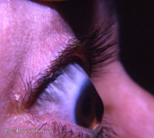
Keratoconus
“File:Keratoconus21.jpg” by William Charles Caccamise, Sr, MD is licensed under CC BY-SA 4.0

Common ophthalmologic considerations for EDS include myopia (near-sightedness), glaucoma and retinal detachment. Another common ocular complication of EDS is keratoconus-a condition in which the cornea bulges outward and then gravity pulls it downward causing blurred vision that isn’t fully corrected even with glasses or contact lenses. This can be treated with rigid contact lenses and various surgical options. Blue sclera may occur in patients with EDS, this is a blue appearance of the whites of the eyes caused by thinness of the sclera. Angioid streaks, generally harmless in themselves, indicate connective tissue disorder as they are breaks in one of the connective tissue layers of the eye and should be monitored for abnormal growth of blood vessels in these cracks. Redundant or drooping skin of the eyelid may be present due to eyelid laxity.4 Dry eyes are also very common in patients with EDS affecting 63% of patients. This may be due to impaired closure of the eyelids resulting in overexposure of the eyes to the environment.6 This dryness can then contribute to another common EDS-related ocular issue- light sensitivity, known as photophobia. Strabismus or abnormal alignment of the eyes either toward a cross-eyed alignment or to the opposite away from midline alignment is another common and not well understood EDS-related issue. This too can be surgically corrected, but surgery is not a good choice for first-line treatment for patients with EDS due to the complications it causes with healing.7 

People with EDS should see an optometrist or ophthalmologist for a comprehensive baseline and exam and then annually for follow-up. It is important to have a relationship with a professional who can determine if any issues that come up are EDS-related and treat them accordingly. Problems that would require intensive evaluation in a patient with EDS include blurred vision or double vision that comes and goes, complete/almost complete vision loss in one eye, migraine auras, dry eyes, light sensitivity, tunnel vision, and floaters. Emergency assistance should be sought if a person with EDS experiences double vision that starts suddenly, flashes of light, pain, redness or discharge, curtain coming up over vision, frontal headache in which patient “hears pulse in the temple,” or sudden change in vision.3 These issues can indicate vascular damage and increased risk of life-threatening arterial rupture in patients with vascular types of EDS.


I help many patients with EDS by giving them tools to manage and treat their musculoskeletal complaints. I am not an expert in vision therapy or eye care. This blog article is meant to be purely informational. If this subject interests you I recommend you check out the references below. As stated above, please consult with an eye care professional for more information about how these issues may affect you.


  1. https://sciencing.com/7-types-connective-tissue-8768445.html
  2. Mao, Jau-Ren and James Bristow. The Ehlers-Danlos syndrome: on beyond collagens. J Clin Invest. 2001 May 1; 107(9): 1063–1069. 
  3. Opthalmology Medical Resource Guide. Ehlers-Danlos National Foundation. 
  4. https://totaleyecare.com/ocular-complications-ehlers-danlos-syndrome/
  5. https://www.chronicpainpartners.com/types-of-eds/
  6. https://www.theraspecs.com/blog/ehlers-danlos-syndrome-light-sensitivity/
  7. James Kundart OD, MEd, FAAO, FCOVD-A. Ehlers Danlos Syndrome and the Eye.

Written by Mindy Nagel, PT, DPT, OCS · Categorized: EDS, Hypermobility

Apr 21 2020

What is evidence-based Pilates?

Joseph Pilates believed that the modern lifestyle, poor posture and inefficient breathing strategies were the roots of poor health when he was developing and solidifying his approach back in the 1950s. If anything, our lifestyle has become even more unhealthy since his time with even more convenience food items, sedentarism, and full-time jobs dedicated to desk work.

Pilates exercises focus on the use of the core to support the spine and provide better support and alignment for the entire musculoskeletal system. The emphasis is on bringing the full attention to the movement so that each exercise can be performed with the best possible precision and control. Breath is used in coordination with core stabilization and movement. This same philosophy of movement aligns well with Dynamic Neuromuscular Stabilization (DNS) according to Prof. Pavel Kolar PT, PhD and the Prague School of Rehabilitation, which emphasizes the relationship between the postural stabilizing function of the diaphragm, breathing, and developmental neurokinesiology.

Pilates’s original system focused on a drawing in of the abdominal muscles to activate the transverse abdominis. Research has shown this to be a less effective way of stabilizing the spine that can actually create a structural weak point at the focus of the drawing in. DNS focuses on eccentric use of all of the abdominal musculature circumferentially creating an outward pressure on the walls of the abdomen together with the downward pressure exerted by the diaphragm. The result is what is called intra-abdominal pressure (IAP). Proper IAP generation results in a stabilizing and decompressive force on the spine.

When I teach Pilates, I combine traditional Pilates principles with these effective stabilization strategies of DNS. This use of the best strategies we currently know from research evidence in combination with the exercises and theories of Joseph Pilates is known as evidence-based Pilates.

Resources:
https://rehabps.com/REHABILITATION/DNS.html
https://www.pilates.com/education/movement-principles

Written by Mindy Nagel, PT, DPT, OCS · Categorized: DNS, Pilates

May 17 2019

Benefits of Dynamic Neuromuscular Stabilization (DNS)

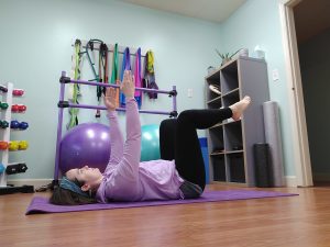
DNS exercise: Basic 3 month supine
DNS exercises use developmental positions to re-establish and strengthen healthy muscle activation patterns.

Dynamic Neuromuscular Stabilization (DNS) is a manual and rehabilitative approach to optimizing the body’s movement system based upon the scientific principles of developmental kinesiology (DK). The developer of DNS is Professor Pavel Kolar, PT, PhD, a Czech physiotherapist and PhD in Pediatrics who has been influenced by the “greats” of the Prague School of Manual Medicine, including Karel Lewit, Vladimir Janda, Vaclav Vojta, and Frantisek Vele.1

[Read more…] about Benefits of Dynamic Neuromuscular Stabilization (DNS)

Written by Mindy Nagel, PT, DPT, OCS · Categorized: DNS

May 06 2019

Hypermobility and proprioception

Did you know you have a sixth sense? No, I’m not talking about ESP. I’m talking about proprioception. Proprioception is the sense of where you are in space. If you close your eyes and I place your right arm in a position out to your side, most people can relatively match that position with their left arm without opening their eyes to look to see exactly where I’ve placed their right arm. This sense of where you are in space helps you control your balance and posture as well. It lets you know if you are fully upright against gravity and helps you maintain yourself upright. In addition, it helps you feel your joint angles and control the alignment of your body in relation to the ground.

[Read more…] about Hypermobility and proprioception

Written by Mindy Nagel, PT, DPT, OCS · Categorized: EDS, Hypermobility, Proprioception

  • « Go to Previous Page
  • Page 1
  • Interim pages omitted …
  • Page 3
  • Page 4
  • Page 5
  • Page 6
  • Go to Next Page »
College Hill Pilates and Physical Therapy LLC
5838 Hamilton Ave.
Cincinnati, OH 45224

Client Portal  •  Terms and Conditions of Use

© 2026 College Hill Pilates and Physical Therapy LLC. All rights reserved.